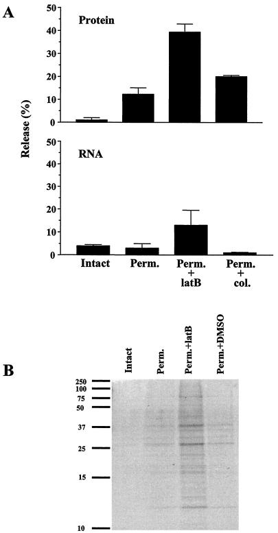
FIG. 4.
(A) Release of protein and RNA from permeabilized cells. Cells were labeled, harvested, and permeabilized as described in Materials and Methods. Acid-precipitable radioactivity present in supernatant and pellet fractions was measured in a scintillation counter. The activity present in the supernatant fraction divided by the total activity in the intact cell is indicated as percent leakage. The upper panel shows the results for the release of 3H-labeled protein, and the lower panel shows the results for the release of 3H-labeled RNA. The values shown represent the averages of two experiments. (B) SDS-PAGE analysis of protein release. Supernatant fractions from intact cells, permeabilized cells (Perm.), permeabilized cells treated with latrunculin B (Perm.+latB), and permeabilized cells treated with DMSO (Perm.+DMSO) were analyzed. A 1/20 volume of each supernatant fraction was fractionated on a 14% polyacrylamide gel. Bands were visualized with Coomassie blue. The positions of prestained broad range protein markers (Bio-Rad) are shown on the left side of the panel.

