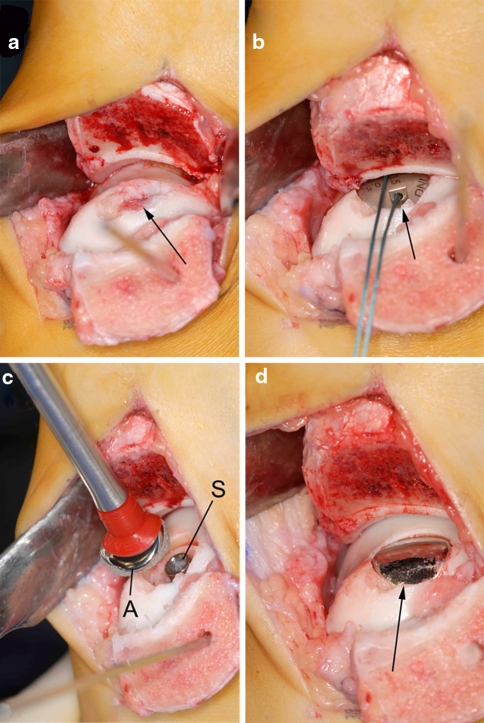
Fig. 2.
Intraoperative pictures of the operative technique. a The talar lesion was exposed through an oblique medial malleolar osteotomy, and the necrotic fragment was excised (arrow). b After the insertion of a screw and the determination of the appropriate offset sizes, a trial articular component (arrow) was placed. c The final articular component (A) was orientated in the correct plane and placed on the screw (S). d Final view of the talus after engagement of the articular component (arrow). Note that the edges of the implant are slightly recessed compared to the adjacent cartilage level

