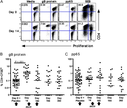
Figure 2.
CD4+ T cells proliferate in response to glycoprotein B (gB) protein. Peripheral blood mononuclear cells (PBMCs) were stimulated in vitro with gB protein, a pool of peptides from cytomegalovirus pp65, or staphylococcal enterotoxin B (SEB). A, The frequency of proliferating CD3+CD4+ T cells was determined by labeling PBMCs with 5, 6-carboxyflourescein diacetate succinimidyl ester (CFSE) at prevaccination (day 0) and 2 weeks after vaccination (day 14) and is shown for a selected individual. Percentage of CFSElo cells is indicated in the upper left hand quadrant. CD4+ T cell proliferative responses are shown in response to gB protein (B) or pP65 protein (C) at the indicated time points. Data for all vaccine-naive subjects (placebo recipients and individuals at the prevaccination time point of day 0) are combined. Net responses (ie, with unstimulated responses subtracted) are plotted. The median response is shown by the horizontal line. Vaccination time points are indicated by an arrow.

