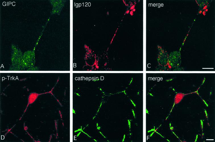
Figure 10.
Endogenous GIPC and TrkA do not colocalize with lysosomal markers. Immunofluorescence staining for endogenous GIPC (A) shows little overlap with that of lgp120 (B), because virtually no overlapping yellow signal is seen in the merged image (C). Similarly, TrkA (D) does not codistribute with cathepsin D (E). Double labeling was performed in NGF-induced, differentiated PC12 (615) as in Figure 8 with polyclonal anti-GIPC and mAb anti-gp120 or anti-TrkA (E-6) and polyclonal anti-cathepsin D. Bar, 10 μm.

