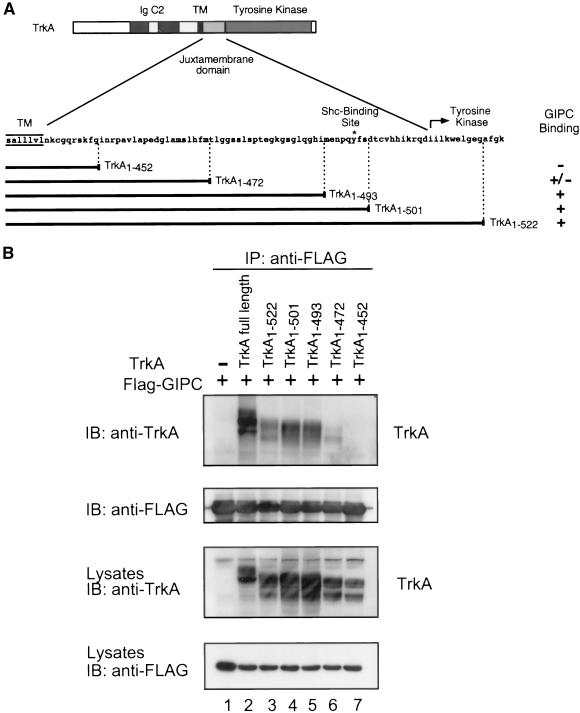
Figure 2.
Mapping the GIPC-interacting region in TrkA. (A) Schematic representation of the structure of TrkA and the juxtamembrane domain deletion mutants. The interaction between GIPC and TrkA mutants shown on the right is based on data from B. (+, interaction; −, no interaction; TM, transmembrane region; IgC2, Ig-like C2-type domains.). (B) Coimmunoprecipitation of GIPC with TrkA deletion mutants. Top panel, full-length TrkA, TrkA1–522, TrkA1–501, and TrkA1–493 coprecipitate with GIPC-FLAG (lanes 2–5), but no interaction is observed with TrkA1–472 or TrkA1–452 (lanes 6 and 7). Second panel, the amount of GIPC-FLAG precipitate is comparable in all lanes. FLAG-tagged GIPC was transiently coexpressed in HEK293T cells with full-length (lane 2) or TrkA deletion mutants (lanes 3–7) or FLAG-GIPC alone (lane 1). Cell lysates were immunoprecipitated (IP) with anti-FLAG followed by immunoblotting (IB) with anti-TrkA RTA (top panel) or anti-FLAG (second panel). Protein expression levels of TrkA mutants (third panel) and FLAG-GIPC (bottom panel) in 50 μg of cell lysate are comparable.

