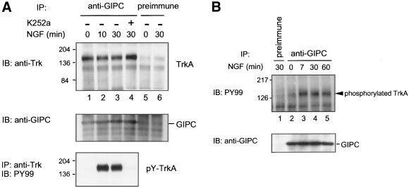
Figure 4.
Association of TrkA with endogenous GIPC in PC12 (615) cells. (A) Top, TrkA coprecipitates with endogenous GIPC in both control (lane 1) and NGF-treated (lanes 2 and 3) cells. Addition of 100 nM K252a, a specific Trk kinase inhibitor, before NGF treatment had no effect on the interaction (lane 4). Little or no TrkA is seen in precipitates obtained with preimmune serum (lanes 5 and 6). Middle, the presence of GIPC in immune (lanes 1–4) but not preimmune precipitates (lanes 5, 6) was verified by immunoblotting. Bottom, an aliquot of lysate was immunoprecipitated with anti-Trk (C-14) IgG and immunoblotted with anti-phosphotyrosine (PY99) IgG to verify the activation of TrkA by NGF (lanes 2 and 3). PC12 (615) cells were cultured in low-serum–containing medium overnight and treated with 100 ng/ml NGF for 0, 10, or 30 min. Cell lysates (3.7 mg) were immunoprecipitated (IP) with either anti-GIPC or preimmune serum followed by immunoblotting (IB) with anti-Trk (B-3) (top) or anti-GIPC (middle). (B) Phosphorylated TrkA (arrowhead) coprecipitated with endogenous GIPC at all time points (7, 30, and 60 min) after NGF treatment (lanes 3–5) but not in the untreated sample (lane 2) or that precipitated with preimmune serum (lane 1). PC12 (615) cells were treated with 100 ng/ml NGF as in A. Cell lysates (3.3 mg) were immunoprecipitated with either anti-GIPC or preimmune serum followed by immunoblotting with antiphosphotyrosine PY99 (top) or anti-GIPC (bottom).

