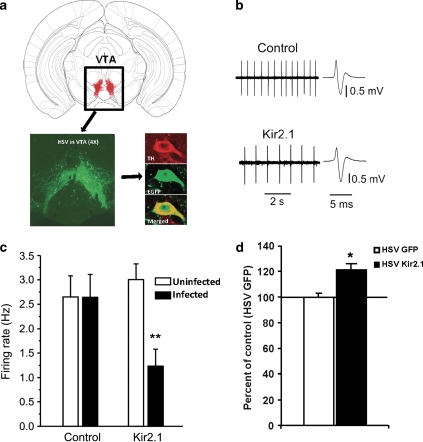
Figure 3.
Expression of herpes simplex virus (HSV)-Kir2.1 decreases the firing rate of dopamine neurons and restores proper cell size in the ventral tegmental area (VTA) of the ClockΔ19 mice. (a) Schematic diagram showing a coronal section of mouse brain containing the VTA, along with a representative image (bottom left) showing a × 4 magnification of the VTA-specific targeting of HSV by stereotaxic surgery after 4 days. On the bottom right are representative dopamine neurons immunostained with enhanced green fluorescent protein (EGFP) and anti-tyrosine hydroxylase (TH) antibodies and images were merged to see colocalization using a confocal microscope. (b) Sample traces and spikes for extracellular recordings in the VTA slice. (c) HSV-Kir2.1 infection reduces the firing rate of dopamine neurons when compared with both ClockΔ19 animals infected with HSV-GFP and neighboring uninfected neurons in ClockΔ19 animals infected with the HSV-Kir2.1. **P<0.01 (one-way ANOVA, n=7–15 cells from 5 to 7 mice per group). (d) The size of the soma of dopamine cells of ClockΔ19 mutant mice injected with HSV-Kir2.1 in the VTA are larger than the cells of mutants that are injected with HSV-GFP (**P<0.01 by t-test, n=4–5 mice per group, 25–35 cells per group).

