Figure 3.
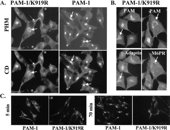
PAM-1/K919R cells: localization and internalization of PAM antibody. (A) AtT-20 cells expressing PAM-1/K919R or PAM-1 were visualized simultaneously with rabbit polyclonal antiserum to PHM (JH1761) and mouse monoclonal antibody to PAM-CD. Bold arrows mark the TGN region and thin arrows indicate sites of vesicular staining and asterisks mark tips of processes. (B) AtT-20 cells expressing PAM-1/K919R were visualized simultaneously with antisera to exon 16 and γ-adaptin or mannose 6-phosphate receptor (M6PR). Arrows mark identical sites in the TGN region. The images shown are representative of the staining patterns observed in multiple independently derived cell lines. (C) AtT-20 cells expressing PAM-1/K919R or PAM-1 were incubated in antibody to exon 16 for 10 min. Antibody was removed and cells were rinsed and chased for 5, 30, or 70 min before fixation and incubation with FITC-tagged goat anti-rabbit immunoglobulin. Identical results were obtained with two additional, independent PAM-1/K919R cell lines.

