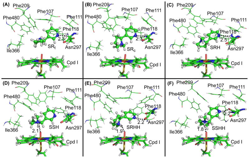
Figure 1.
Representative CYP2A6-(S)-(−)-nicotine binding structures derived from the trajectory of MD simulations. Cpd I of CYP2A6 is shown in stick style, and residues within 5 Å around the ligand are shown in lines. (S)-(−)-nicotine is displayed in ball-and-stick style. Possible hydrogen bonds between CYP2A6 and (S)-(−)-nicotine are represented by dashed lines with the distances (Å) labeled. A: CYP2A6-SRt; B: CYP2A6-SRc; C: CYP2A6-SRH; D: CYP2A6-SSH; E: CYP2A6-SRHH; and F: CYP2A6-SSHH.

