Figure 2.
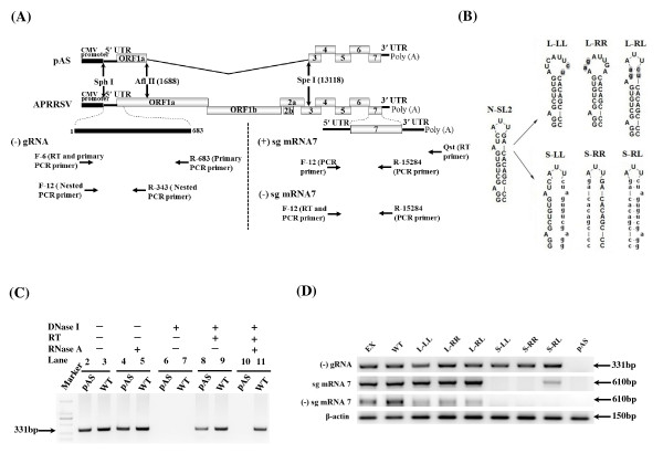
Mutational analysis of the predicted stem-loop structure in the N-SL2. (A) Strategic representation of RT-PCR used to detect (-) gRNA, (+) sg mRNA7 and (-) sg mRNA7. The positions are according to APRRSV stain (GenBank: GQ330474) and all primer sequences are listed in Table 1. pAS was a non-replicative control which was absence of gene ORF1a and ORF1b (1688-13118) in full-length cDNA clone. (B) Schematic representation of the mutations introduced into the N-SL2 structure. The loop was enlarged as described in Figure 1, and mutants L-LL and L-RR were generated by overlapping PCR mutagenesis. L-RL was generated by combining the right and left arm sequences of the L-LL and L-RR, respectively, such that the overall structure of N-SL2 was restored. All the mutated nucleotides (lowercase) are highlighted in gray shading. The stem mutants, S-LL and S-RR, were generated by overlapping PCR such that one arm sequence was replaced with that of the opposite arm. The double mutant, S-RL, was generated by combining the mutations in the left and right arms such that the overall structure was restored. All mutant sequences are shown as lowercase. (C) RT-PCR of RNAs extracted from pAS and WT transfected cells at 24 hours after transfection. DNase I and RNase A were used to omit template DNA and the reverse transcriptase. The primers were nested RT-PCR primers as same as (-) gRNA detection. A 2-kbp ladder was used as a molecular size marker. The numbers indicated the lane No. (D) RT-PCR analysis of the mutants. Total cellular RNAs were extracted from mutant plasmids-transfected from BHK-21 cells at 24 hours post-transfection. β-actin is a marker for the level of intracellular RNA isolation, and pAS is a non-replicative control.

