FIG. 4.
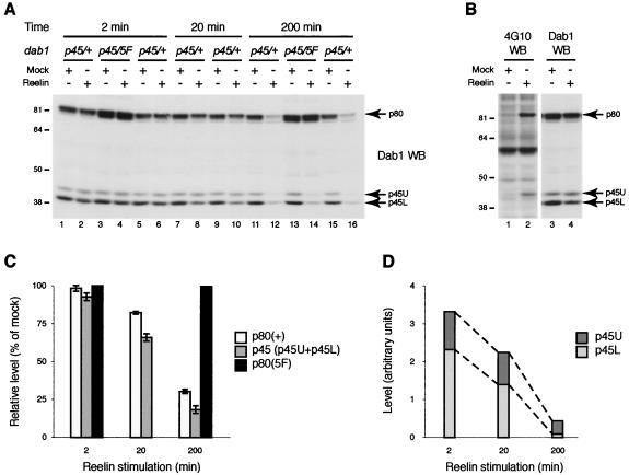
Reelin-induced tyrosine phosphorylation of Dab1 is necessary for its degradation. (A) Cortical neuron cultures were prepared from littermate embryos derived from a cross between a dab15F/+ female and a dab1p45/p45 male and subsequently genotyped. Each culture was treated with Reelin-containing or mock supernatant (as indicated) for 2 (lanes 1 to 6), 20 (lanes 7 to 10), or 200 (lanes 11 to 16) min. Total lysates were subjected to SDS-PAGE and Western blot analysis using anti-Dab1 antibodies (Dab1 WB). The respective positions of full-length Dab1 (p80), Dab1 p45 of slower electrophoretic mobility (upper band [p45U]), and Dab1 p45 of faster mobility (lower band [p45L]) are indicated by arrows. (B) Neuron cultures prepared from a dab1p45/+ embryo were treated with Reelin-containing (lanes 1 and 3) or mock (lanes 2 and 4) supernatant for 20 min. Total lysates were subjected to SDS-PAGE and Western blot analysis using anti-Dab1 antibodies to detect total Dab1 (Dab1 WB) and an anti-phosphotyrosine antibody to detect tyrosine-phosphorylated Dab1 (4G10 WB). The respective positions of full-length Dab1 (p80), Dab1 p45U, and Dab1 p45L are indicated by arrows. (C) Quantification of data presented in panel A. Relative levels of different Dab1 forms, wild-type Dab1 [p80(+)], Dab15F [p80(5F)], and total Dab1p45 [p45 (p45U+p45L)] after Reelin stimulation for 2, 20, or 200 min. The values are presented as percentages of the average level of each form in mock-treated samples. Error bars represent standard deviations. (D) Quantification of data presented in panel A. Relative levels of Dab1 p45U and Dab1 p45L after Reelin stimulation for 2, 20, or 200 min are shown. The values are presented as densitometric units.

