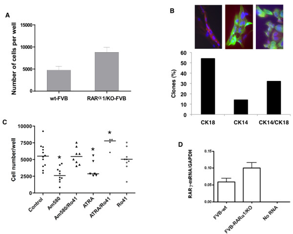
Figure 3.

Growth, composition and regulation of primary mammospheres. (a) Comparison of mammosphere growth. Primary cells obtained from wt and RARα1/KO mammary glands, were inoculated at 2.5 to 5 × 10⁴ under conditions of ultra-low adhesion and serum-free medium and eight days later, the mammospheres were disassociated and the cells counted (see Materials and methods). The bars show mean (4,750 and 8,833 cells respectively, and SD of three individual experiments and three to four samples per group; P = 0.04 by unpaired t-test. (b) Bi-potential cells in mammospheres. Primary mammary epithelial cells were incubated for eight days until most single cells died; mammospheres were dissociated, inoculated into 96 wells, at 1cell/well (see Materials and methods) and when colonies formed stained for CK18 and CK14. (Red - CK18; green - CK14; blue - DAPI). Total number of colonies stained = 21. Scale bar = 10 um. (c) Regulation of mammosphere growth by retinoids. Cells isolated from mammary glands of seven- to eight-week old FVB mice were inoculated under conditions of mammosphere formation and eight to ten days later were treated with 10 nM of Am580, 200 nM of atRA, and/or 10-fold excess (100 nm and 2 μM, respectively) of RARα antagonist, Ro41-5253 for seven to eleven days. Each symbol represents an individual well; there were two to four wells per experiment, and a total of four experiments. Kruskal-Wallis test P = 0.0002; *indicates significance at P < 0.05 by Dunn's Multiple Comparison Test. (d) RARγ1 expression. RNA was extracted from freshly isolated and partially purified mammary epithelial cells and wt and RARα1/KO, and subjected to Q-PCR analysis as described in Materials and methods. The bars show mean of three experiments, two samples in each.
