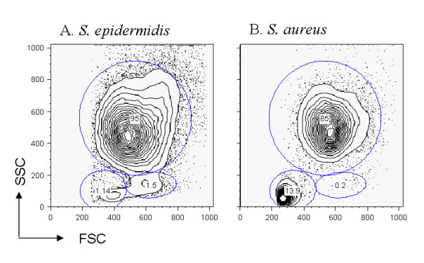
Figure 2.

Cell population in the milk after S. epidermidis and S. aureus challenges. After incubation with propidium iodide, cells from cisternal lavages were analysed by flow cytometry. Dead cells were electronically gated out, and cell types (granulocytes, monocytes/macrophages and lymphocytes) were analysed on the forward and side scatter intensity profiles. The results from a resistant ewe after Se (A) or Sa (B) are presented.
