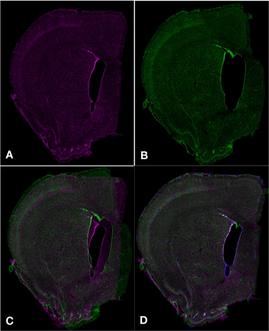
Figure 5.
Aligning serial sections using Mogrifier. Two serial coronal sections (A,B) are shown as they would appear in Mogrifier (i.e., in red and green channels). Image (B) illustrates an example of non-uniform compression of the lateral ventricle in a tissue section. Image (C) shows the overlay of slices (A,B) in the application Mogrifier before applying any transformation. Image (D) shows the overlay of slices (A,B) and the reference points (blue) after applying the thin plate spline transformation in Mogrifier.

