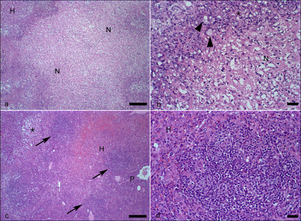
Figure 2.

Histology of liver from hamsters. Histology of liver from hamsters inoculated with the HM1 strain of E. histolytica. (a) significant necrosis of the liver parenchyma (N) of untreated hamster melatonin. Non-necrotic hepatic parenchyma (H), (b) Detail of preceding figure showing trophozoites (arrowheads) adjacent to the large amount of cellular debris and moderate inflammatory infiltrate (*). Necrosis (N), (c) non-necrotic hepatic parenchyma (H) of hamsters that received melatonin. Note the presence of focal inflammatory infiltrate rich in mononuclear cells (arrows). Granulation tissue (*). Portal space (P), (d) Detail of previous picture showing one of the focal infiltrates predominantly composed of lymphocytes and macrophages. Non-necrotic liver parenchyma (H). H&E. (a) and (c) Bar 100 μm; (b) and (d) Bar 20 μm.
