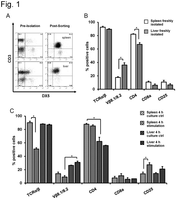
Figure 1.
FACS analysis of T cell marker from spleen and liver CD3+/DX5+NKT cells in Balb/c mice. DX5+NKT cells were isolated from spleen and liver of Balb/c mice using MACS and FACS-Sorting. Representative dot plot (A). Expression of different T cell markers revealed distinct subsets in Balb/c mice. In the spleen, more DX5+NKT cells appeared CD4+, in the liver, they expressed more Vβ8.1/8.2 (B). Upon 4 h activation with anti-CD3 and anti-CD28, spleen DX5+NKT cells displayed a TCRα/β down-regulation, whereas liver DX5+NKT cells did not (C). Results are given as mean + SEM. Experiments were repeated at least three times (* p < 0.05).

