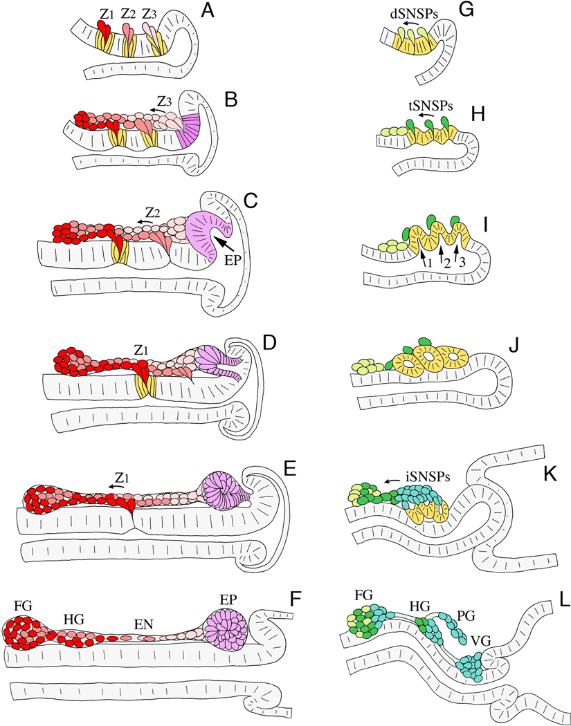
Figure 3. Schematic illustration of neurogenesis in Manduca (A–F; after Copenhaver and Taghert, 1991) and Drosophila (G–L; after Hartenstein, 1997).
Each panel represents a sagittal view of the foregut midline; anterior is to the left, dorsal is to the top. A. At ~24% of development in Manduca, three neurogenic zones have formed in the dorsal foregut epithelium (Z1, Z2, & Z3), from which a series of individual precursors will delaminate; each precursor cell divides once or twice after emerging. B. By 28% of development, streams of zone-derived cells have begun to migrate anteriorly along the foregut, while all of the remaining zone 3 cells delaminate. The epithelium surrounding the original position of zone 3 subsequently differentiates into a distinct placode (purple). C. By 33% of development, migrating zone cells have begun to form the frontal ganglion anteriorly, while all of the remaining zone 2 cells delaminate. The EP cell placode has also begun to invaginate. D. At 36% of development, zone-derived cells continue to migrate along the pathways that will form the recurrent and esophageal nerves, while the EP cell placode invaginates. E. By 39% of development, the last of the zone 1 cells delaminate, and the EP cell placode has invaginated onto the foregut surface. F. By 42% of development, the frontal and hypocerebral ganglia have formed; subsets of the residual zone cells will proliferate to form glial populations that ensheath the nerves and ganglia of the ENS. At this stage, the invaginated EP cells form a condensed packet of post-mitotic neurons adjacent to the foregut-midgut boundary. G. In Drosophila at stage 10 (~24% of development), three neurogenic centers form within the ENS anlage (yellow) and give rise to a set of delaminating precursors (dSNSPs; light green). H. At stage 11 (~30% of development), the dSNSPs have migrated anteriorly, where they will help form the frontal ganglion and its nerves. A second wave of precursors (tSNSPs, dark green) delaminates from the neurogenic centers, marking the positions where three invaginations will form. I. By stage 12 (~34% of development), the three invaginations form distinct pouches (1, 2, & 3) that protrude onto the foregut surface. J. By stage 13 (~38% of development), the three pouches have pinched off to form a set of neurogenic vesicles, while the tSNSPs migrate anteriorly to help form the frontal and hypocerebral ganglia. K. By stage 14 (~42% of development), invaginated cells from the three vesicles begin to dissociate (iSNSPs) and migrate anteriorly. L. By stage 16 (~60% of development), the SNSPs have assembled into the enteric ganglia of the foregut: frontal ganglion (FG), hypocerebral ganglion (HG), paraesophageal ganglion (PG), and ventricular ganglion (VG). Processes from these neurons also pioneer the interganglionic nerves.

