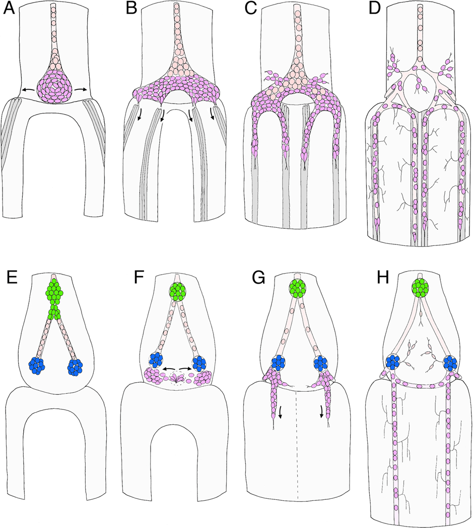
Figure 6. Schematic illustrations of the sequence of migration that forms the midgut enteric plexus in the insect ENS.
A–D represents Manduca (after Copenhaver and Taghert, 1989b; Copenhaver, 1993); E–H represents Schistocerca (after Ganfornina et al., 1996). All panels show dorsal views of the developing ENS at the foregut-midgut boundary. A. At 40% of development, the EP cells have invaginated onto the dorsal foregut surface and begin to spread bilaterally around the foregut-midgut boundary. Concurrently, subsets of longitudinal muscles on the midgut (dark grey cells) begin to coalesce as dorsal closure of the midgut proceeds. Anteriorly, the EP cell packet is in continuity with the residual zone 3 cells that help form the esophageal nerve; subsets of these zone-derived cells will subsequently proliferate to form glial cells that ensheath the enteric plexus. B. By 55% of development, the EP cells have almost completely surrounded the foregut, and subsets of the cells have begun to align with each of the eight midgut muscle bands (only the dorsal four are shown). C. By 58% of development, the EP cells have begun to migrate posteriorly along the muscle bands on the midgut; a small number of neurons also migrate laterally onto radial muscles of the foregut (foregut muscles not shown). D. By 80%, the EP cells have completed their migration, forming the enteric plexus that spans the foregut-midgut boundary; they have also extended axons along the posterior midgut (not shown) and short terminal branches onto the adjacent interband musculature. Glial cells (pink) derived from the residual zone 3 cells have also migrated over the major branches of the enteric plexus to ensheath them. E. By 40% in the Grasshopper ENS, the neurogenic zones of the foregut have given rise to the cells of frontal ganglion (not shown) and hypocerebral ganglion (green), while posteriorly, cells derived from the third neurogenic zone have migrated bilaterally to form the incipient ingluvial ganglia (blue). F. By ~50% of development, a second wave of neurogenesis from the vicinity of zone 3 has begun to produce a new population of ingressing cells (purple); these cells then migrate bilaterally and aggregate adjacent to the ingluvial ganglia. G. By ~60% of development, four streams of cells have begun to migrate posteriorly from the ingluvial ganglia onto the midgut (only the dorsal pair is shown). Unlike Manduca, distinct muscle bands on the midgut have not been detected in grasshopper. H. By 80% of development, the migratory populations of neurons have become distributed along the entire length of the midgut and have extended terminal branches onto the adjacent musculature. An additional set of cells derived from the neurogenic zones (presumably sensory neurons) also contributes to an extensive foregut plexus (not detected in Manduca).

