
Fig. 1.
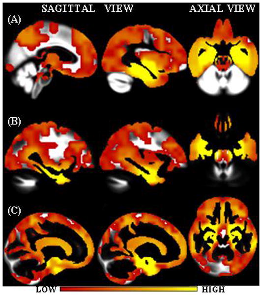
Interpolated weight vectors for (A) GM, (B) WM and (C) CSF overlaid on the corresponding custom template. Color scale indicates the weight i.e. the importance of the voxel location for classification.
Official websites use .gov
A
.gov website belongs to an official
government organization in the United States.
Secure .gov websites use HTTPS
A lock (
) or https:// means you've safely
connected to the .gov website. Share sensitive
information only on official, secure websites.
Interpolated weight vectors for (A) GM, (B) WM and (C) CSF overlaid on the corresponding custom template. Color scale indicates the weight i.e. the importance of the voxel location for classification.