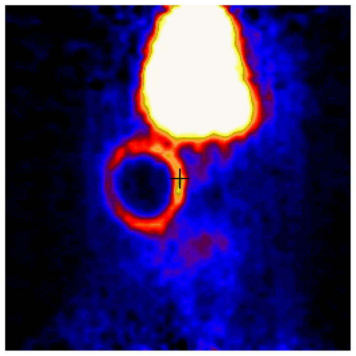
Figure 2. Representative coronal plane of abdomen of Lewis Rats transplanted with allogeneic islets.

Rats were imaged (90 min) dynamically with 250 uCi [11C]DTBZ and a Concorde microPET scanner. The large high uptake area in the top center of the figure is a plane of the liver, an organ of [11C] DTBZ catabolism. Radioligand uptake in the form of a ring corresponds to the location of the transplanted islets. Following imaging, the presence of insulin staining cells in this location was confirmed by preparing paraffin embedded sections of the tissue and insulin staining by immunohistochemistry.
