
Figure 11.
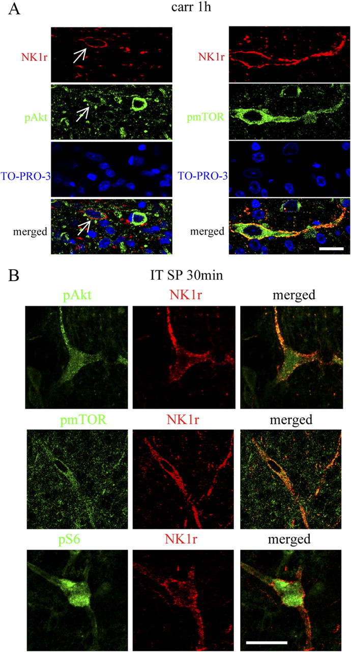
pAkt, pmTOR, and pS6 are colocalized with NK1R in laminae I, III, and IV. A, Confocal images of pAkt or pmTOR (green) and NK1R (red) at 1 h after carrageenan. In each case, a single NK1R-labeled neuron is seen also pAkt or pmTOR positive. Sections are counterstained with TO-PRO-3 (blue) to label cell nuclei. B, Confocal images of pAkt, pmTOR, or pS6 (green) and NK1R (red) at 30 min after intrathecal SP (30 nmol). A large lamina III/IV neuron is shown. NK1R internalization can be seen on the dendrites and soma. Single focal plane images are shown. Scale bars, 20 μm.