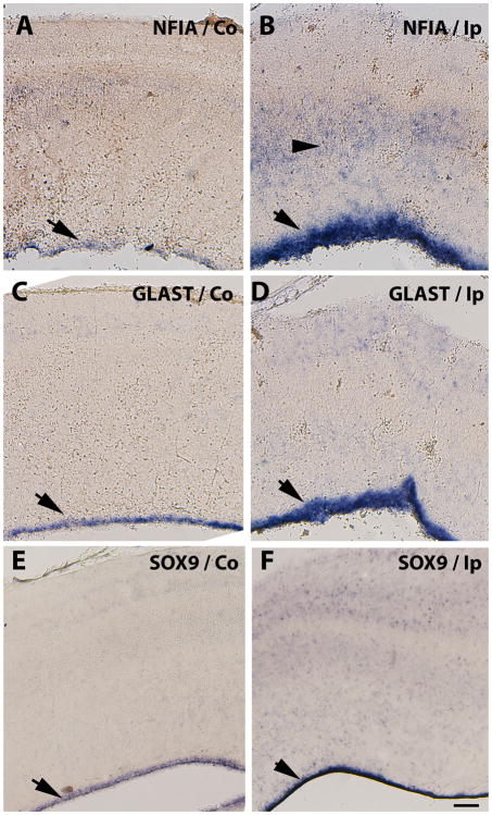
Figure 6. Increased expression of astrocytic precursor markers in the VZ of embryos injured at E11.
Detection of NFIA (A, B), GLAST (C, D) and SOX9 (E, F) mRNAs in serially adjoining cross-sections of the tectal laminae three days after stab injuries performed at E11. Increased expression levels of astrocyte precursor markers in Ip (ipsilateral) injured tecta (B, D and F) with respect to the Co (contralateral) side (A, C and D) in the VZ (arrows) and scattered throughout the tectal laminae (B arrowheads) suggests activation of astrocyte differentiation. Scale bar: 100μm. Results depicted in this figure were consistently observed in multiple independent embryos (3 embryos).

