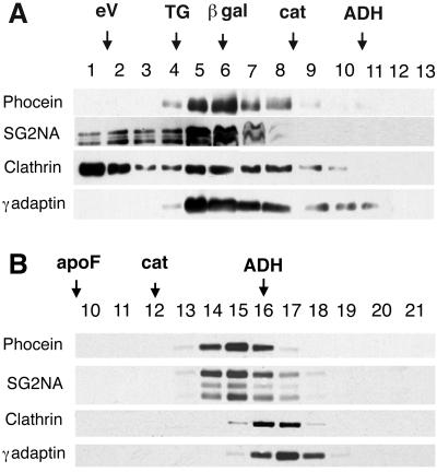
Figure 4.
Gel filtration and sucrose gradients. (A) Cytosolic brain proteins were gel filtered as described in MATERIALS AND METHODS. Fractions 1 and 2 are the excluded volume eV. (B) Cytosolic brain proteins were overlaid on sucrose gradients as described. Aliquots of the fractions were analyzed by Western blotting and revealed with the indicated antibodies. Calibrating proteins are indicated by arrows above the fraction numbers: porcine thyroglobulin, TG; β-galactosidase, β-gal; apoferritin, apo F; catalase, cat; and alcohol dehydrogenase, ADH.

