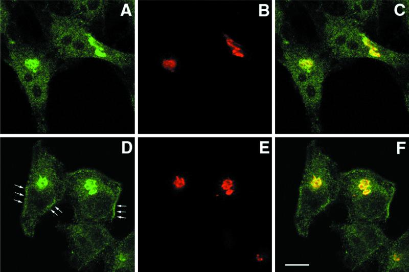
Figure 6.
Phocein and SG2NA localize to the Golgi apparatus in HeLa cells. HeLa cells were fixed, permeabilized, and processed for immunofluorescence microscopy using rabbit, affinity-purified anti-phocein (A) and anti-SG2NA antibodies (D) and a mouse monoclonal antibody raised against CTR433 (B and E), revealed by an Alexa 488-labeled antibody raised against rabbit immunoglobulins and a Cy5-labeled antibody raised against mouse immunoglobulins. Cells were observed under a confocal microscope. Medial optical cuts of representative cells are shown (C and F). Areas of colocalization appear yellow in the computer-generated composite image. Bar, 10 μm.

