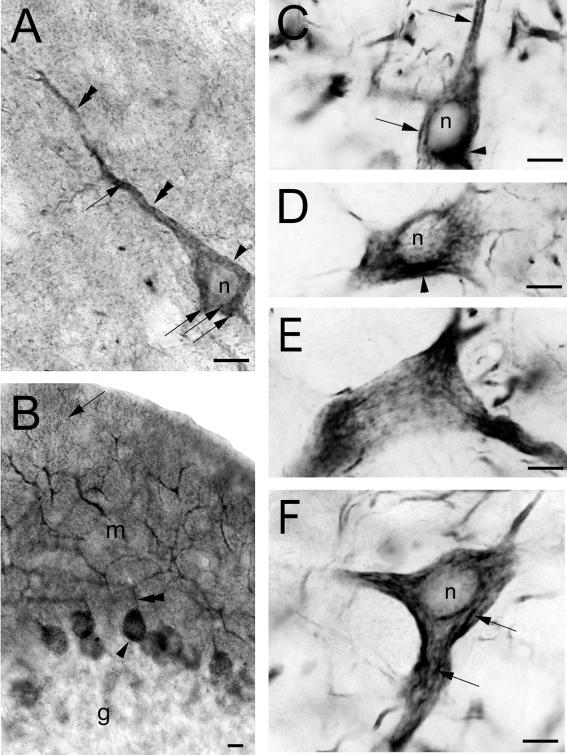
Figure 8.
Somato-dendritic localization of phocein in neurons. (A) Phocein immunoreactivity is restricted to the soma (arrowheads) and dendrites (double arrowheads) of cortical pyramidal cells. The intracytoplasmic labeling present in the soma and apical dendrite is granular (arrows). (B) Phocein labeling is present in the soma (arrowhead) and all branches of the dendritic arborization of cerebellar Purkinje cells, from the large proximal dendrites (double arrowhead) to the most distal, thin dendrites (arrow). (C–F) Intracytoplasmic, vermiculated labeling (arrows in C and F) is visible in the soma and emerging proximal dendrites (E) of neurons in the spinal trigeminal nucleus (C and D), in the bulbar reticular nucleus (E), and in the red nucleus (F). A labeled area close to the nuclei may correspond to a Golgi apparatus (C and D). In D, a network reminiscent of the ER is labeled. Note that nuclei (n) are devoid of staining. Bars, 10 μm.

