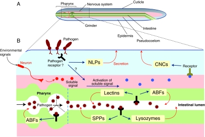
Fig. 1.
C. elegans antimicrobial defences. (A) Basic anatomy of C. elegans. The worm is composed essentially of two concentric tubes – the epidermis (also called hypodermis) and the intestine – separated by a fluid cavity, the pseudocoelom. The physical barriers of the worm include a grinder, which mechanically disrupts the bacteria that form the worm’s normal diet, and the cuticle, which envelops the animal. (B) Selected AMPs and proteins in C. elegans. Invading microbes induce cell damage and are also believed to be detected via specific pathogen receptors. These two events trigger specific signalling pathways that lead to the production and release of a variety of specific antimicrobial proteins and AMPs (yellow boxes). Depending on the pathogen, these antimicrobial effectors are expressed specifically in a particular tissue, such as the pharynx, the intestine or the epidermis. Figure adapted with permission (Millet and Ewbank, 2004).

