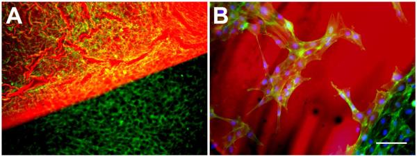
Figure 1. In vitro cytocompatibility.
After 48 hrs exposure to the adhesive complex coacervate, osteoblast viability (live/dead) and cell morphology (phalloidin-FITC staining) assays showed cell contact spreading and proliferation. A) Adhesive associated with osteoblast attachment on tissue culture polystyrene, showing homogeneous staining both on the polystyrene and the adhesive. B) Magnified view from A, showing osteoblast attachment directly to the adhesive. Adhesive (red), actin (green), and DAPI (blue). Scalebar = 100 μm.

