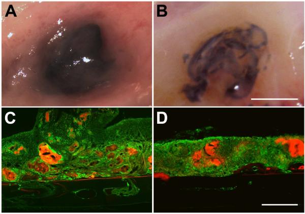
Figure 3. Analysis of the adhesive on skull surface.
2 weeks (A,C) following the application of 1 μl of adhesive directly on the surface of the skull, the adhesive was found to be largely intact. Skull sections showed CD68 immunoreactivity (green) associated with adhesive fragments (red). Adhesive degradation was more obvious at 4 weeks (B,D). A and B show representative views of the adhesive and associated tissue at the time of sacrifice. C and D show representative sagittal skull sections through the center of the adhesive droplet. Sections are oriented with the side in contact with the dura down and the nose of the animal to the left. Scalebar in B, same as A, and = 5 mm. Scalebar in D, same as C, and = 500 μm.

