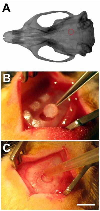
Figure 4. Rat calvarial model.
A) CT reconstruction of rat skull, with location of the craniotomy shown in red. B) Rats had a 3 mm diameter burr hole with the center positioned −3.2 mm from bregma, and 2 mm lateral to bregma cut via trephination. C) The craniotomy was replaced with 1 μl of adhesive (n = 8). Additional sham animals had the craniotomy replaced without adhesive (n = 4). Scalebar in C, same as B, and = 5 mm.

