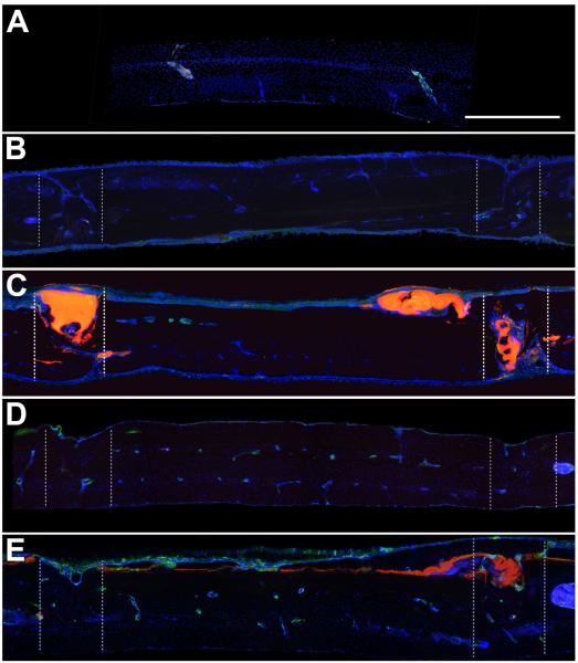
Figure 5. CD68 Immunoreactivity.
A) Representative sagittal control skull section showing minimal CD68 immunoreactivity, which was usually associated with marrow cavities. B) Representative sagittal section from the sham group at 4 weeks, showing fusion of the bones on one side and incomplete fusion on the other. C) Representative sagittal skull section at 4 weeks from the adhesive group showing adhesive within and on the surface of the craniotomy, associated with CD68 immunoreactivity. The adhesive was sufficient to maintain the placement of the craniotomy. Complete bone fusion was not observed on either side of the craniotomy. D) Representative sagittal section from the sham group at 12 weeks, showing fusion of the craniotomy on both sides. E) Representative sagittal skull section at 12 weeks from the adhesive group showing adhesive within and on the surface of the craniotomy, associated with CD68 immunoreactivity. Bone fusion was achieved by 12 weeks. Sections are oriented with the side in contact with the dura down and the nose of the animal to the left. CD68 (green), DAPI (blue), bioadhesive (red). Zones of the craniotomy are shown in dashed lines. Scalebar = 500 μm.

