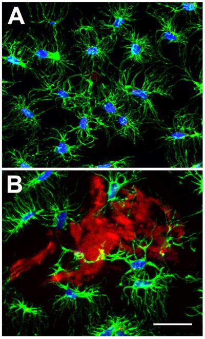
Figure 8. Cellular association with adhesive complex coacervates in vivo.
A) Representative confocal image showing actin (green) and DAPI (blue) staining of osteocytes in normal uninjured rat calvaria. B) Representative confocal imaging showing osteocyte association with adhesive fragments (red) within the craniotomy region at 12 weeks. Scalebar = 50 μm.

