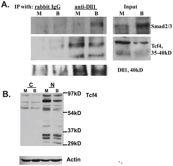
Fig. 2.
Dll1 associates with Smad2/3 and Tcf4 in lysates of HCT-116 CC cells. (A) Immunoprecipitation (IP) analyses with an anti-Dll1 antibody. Nuclear protein lysates (100 μg) from HCT-116 cells, exposed to mock (M) or 5 mM butyrate (B) treatment for 17h, were used for IPs with 2 μg of normal IgG (control) or with 2μg of anti-Dll1 antibody (sc-9102, Santa Cruz Biotechnology). Smad2/3 proteins were detected with a polyclonal antibody that recognizes both proteins (sc-8332, Santa Cruz Biotechnology), Tcf4 was detected with a monoclonal antibody from Millipore (clone 6H5-3). Control experiments confirmed that the anti-Dll1antibody immunoprecipitates the 40kD form of Dll1; we were unable to detect this form in the input, due to its low abundance. (B) Nuclear (N), but not cytoplasmic (C) fractions of HCT-116 cells exhibit the presence of low molecular weight Tcf4 isoforms (30–45 kD). Fractionation of HCT-116 cells exposed to mock (M) or 5 mM butyrate (B) treatment for 24h was performed with the Pierce Nuclear and Cytoplasmic Extraction Reagent Kit (NE-PER). Detection of Tcf4 was carried out as in (A).

