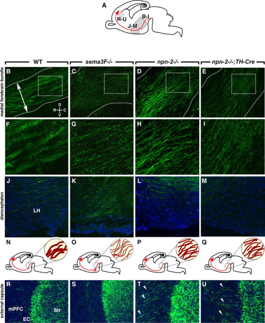
Figure 8.

Abnormal channeling, fasciculation, rostral growth, and EC crossing of mdDA projections in sema3F- and npn-2-deficient mice. A, Schematic showing in gray the derivation of the sections in B–M and R–U. The developing mesoprefrontal projection is shown in red. B–M, R–U, Sagittal sections at the level of the MFB (B–I), diencephalon (J–M), and EC (R–U) of E18.5 wt (n = 5) (B, F, J, R), sema3F−/− (n = 4) (C, G, K, S), npn-2−/− (n = 4) (D, H, L, T), and npn-2−/−;TH-Cre (n = 3) (E, I, M, U) mice. Sections are immunostained for TH (green) to visualize mdDA axons and counterstained with fluorescent Nissl (blue). The boxed areas in B–E are enlarged in F–I. N–Q, Schematic representations of the development of the mesoprefrontal projection (in red) of wild-type (N), sema3F−/− (O), npn-2−/− (P), and npn-2−/−;TH-Cre (Q) mice with an enlargement at the level of the MFB (dark red within circle). B, F, In wild-type mice, the compact MFB (boundaries indicated by dotted lines) projects into the diencephalon with individual TH axons fasciculated into thick bundles. The double-arrowed line in B indicates location of line used to quantify MFB width and axon fascicle number. C, G, In sema3F−/− mutants, the MFB occupies a larger dorsoventral area and individual TH-positive axons are severely defasciculated. In npn-2−/− (D, H) and npn-2−/−;TH-Cre mice (E, I), the MFB is broader and TH-positive axons are defasciculated. In sema3F−/− mutants (K), but not in wild-type (J), npn-2−/− (L), and npn-2−/−;TH-Cre mice (M), many axons in the diencephalon are displaced ventrally and appear to stall in the LH region. R–U, A subset of axons innervates the mPFC by crossing the EC (R). Although this innervation appears normal in sema3F−/− mice (S), TH-positive axons excessively cross the EC in npn-2−/− and npn-2−/−;TH-Cre mice (arrowheads). Str, Striatum.
