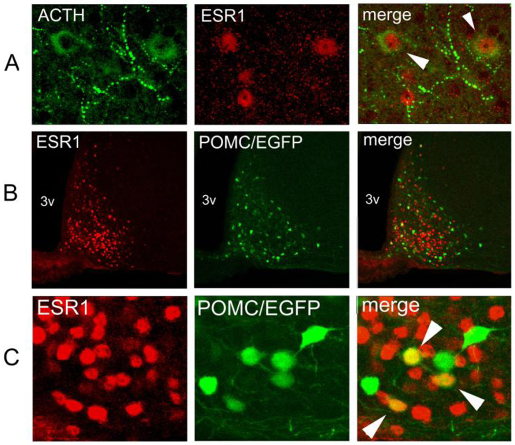
Fig. 4.
Colocalization of estrogen receptor α (ESR1) and POMC in the adult hypothalamus. (A) Immunofluorescence of the rat arcuate nucleus against POMC-derived ACTH (green), ESR1 (red) and the superimposition of both images (merge). Colocalization, denoted by arrowheads, is seen between nuclear ESR1 and cytosolic ACTH. (B) Immunofluorescence against ESR1 (red) on a coronal hypothalamic section of a Pomc-EGFP transgenic mouse (green). 3v: third ventricle. (C) Higher magnification of the same experiment as on B. Colocalization of ESR1 and EGFP is indicated in several neurons with white arrowheads.

