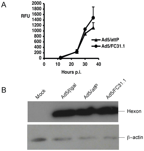
Figure 3. Analysis of protein expression.
HEK-293 cells were infected with Ad5/βgal, Ad5/attP or attB-Ad at MOI of 5, and samples were analyzed at 24, 30 and 36 hpi. Non-viral marker GFP protein was quantified by measuring relative mean fluorescence intensity per cell (RFU/cell) by Flow Cytometry (A). Late viral hexon protein was detected from protein extracts by Western Blot analysis at 24 hpi (B). Equal amount of proteins were loaded and β-actin protein was used as control. Mock are uninfected HEK-293 cells.

