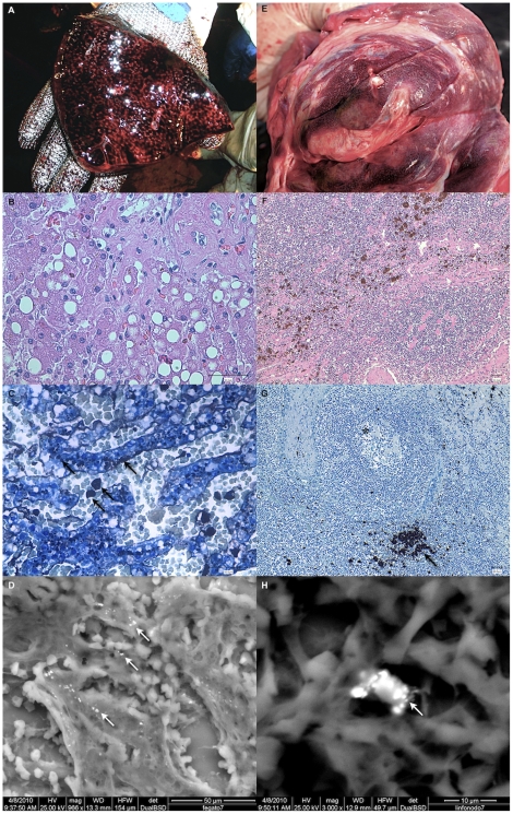
Figure 4. Liver and lymph node pathological changes related to Hg presence.
Gross features, hematoxylin and eosin (HE) and automethallographic staining, along with ESEM pictures of liver (respectively, Fig. A–D; Fig. B and C have a 10× magnification) and lymph nodes (respectively, Fig. E–H; Fig. B and C have a 40× magnification). In HE-stained sections, hepatic macrovacuolar lipidosis in the liver and macrophages loaded with brownish pigments are evident. HgSe crystals can be observed in the liver (hepatocytes and Kuppfer cells) and lymph nodes (sinusal macrophages) both with the automethallographic staining (black arrows) and ESEM pictures (white arrows). In Fig. G also follicular lymphoid depletion is prominent (*).

