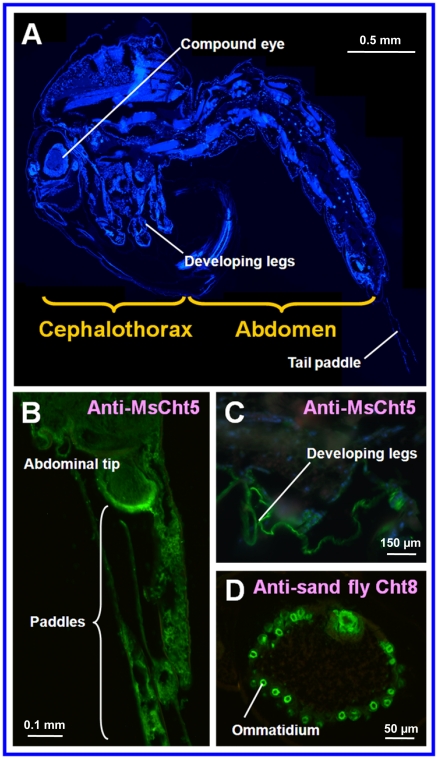
Figure 8. Immunohistochemical localization of selected chitinase proteins expressed in An. gambiae pupae.
A) A paraffin-embedded thin section of a whole pupa showing the overall structure and corresponding regions where chitinases were detected in immunohistochemical analysis as shown in Panels B, C and D. B) Chitinase detected in the abdominal tip and the tail paddles of a pupa by anti-Manduca sexta chitinase 5 polyclonal antibodies (anti-MsCht5) as shown by green color. C) Chitinase detected in certain parts of thorax and developing legs of a pupa by anti-MsCht5 as shown by green color. D) Chitinase detected in the ommatidia of a compound eye by anti-sand fly (Lutzomyia longipalpis) chitinase 8 (anti-sand fly Cht8) polyclonal antibodies as shown by green color.

