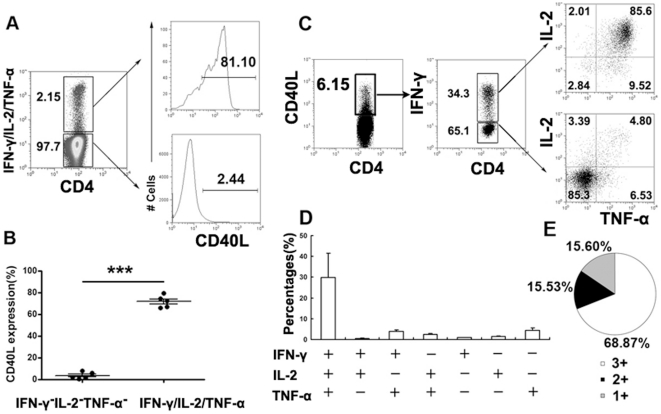
Figure 3. CD4+CD40L+ T cells express polyfunctional cytokines.
PFCs were stimulated with ESAT-6/CFP-10 peptides, anti-CD28 and anti-CD49d mAbs for eight hours. For detection of intracellular cytokines, anti-IFN-γ, anti-IL-2 and anti-TNF-α monoclonal antibodies were labeled with the same fluorescence. The staining of CD40L and cytokines of IFN-γ/IL-2/TNF-α were conducted in one FACS tube.(A) Th1 cytokine (IFN-γ, IL-2 or TNF-α) producing and nonproducing cells were gated. The expression of CD40L was evaluated. (B) Summary of the CD40L expression data within Th1 cytokine producing and nonproducing cells. Horizontal lines represent mean ± SEM. (C) CD4+IFN-γ+ and CD4+IFN-γ− cells within CD4+CD40L+ T cells were further gated, and the expression of IL-2 and TNF-α are shown. (D) The total antigen response within CD4+CD40L+ T cells was defined as the number of cells expressing any combination of IFN-γ, IL-2 or TNF-α. The average percentages for each subset are shown. (E) The percentages of cells producing three cytokines (triple positive), two cytokines (double positive) or only one cytokine (single positive) within the total CD4+CD40L+ T cell response. Data shown are the mean values from five independent experiments.

