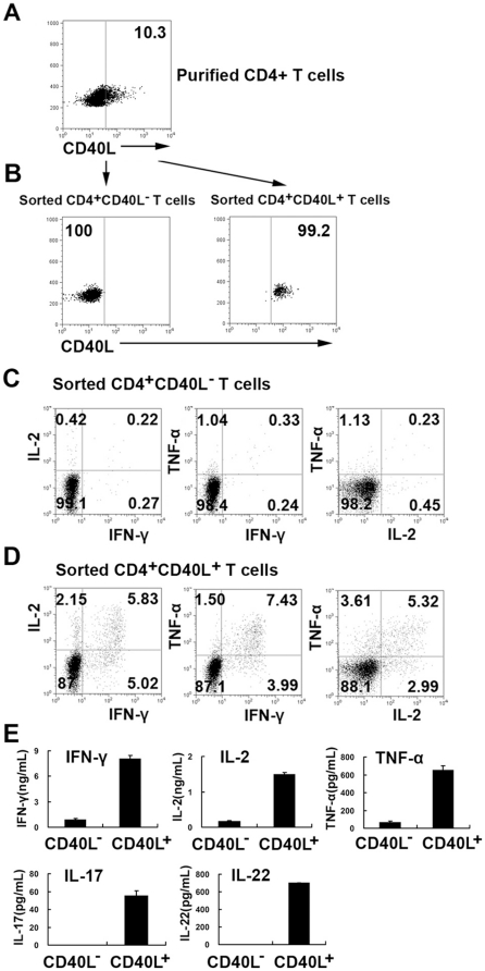
Figure 5. Isolation of viable CD4+CD40L+ and CD4+CD40L− T cells.
PFCs were stimulated with ESAT-6/CFP-10 peptides, anti-CD28 and anti-CD49d mAbs in the presence of a fluorescently labeled anti-CD40L monoclonal antibody and monensin. CD4+T cells were first isolated with magnetic-beads. (A) The expression of CD40L within purified CD4+ T cells was demonstrated, and (B) CD4+CD40L+ and CD4+CD40L− T cells were further sorted by flow cytometry. (C) Sorted CD4+CD40L+ or (D) CD4+CD40L− T cells were co-cultured with purified CD14 cells independently in the presence of ESAT-6/CFP-10 peptides. The intracellular expression of IFN-γ, IL-2 or TNF-α by sorted CD4+CD40L+ and CD4+CD40L− T cells was detected by flow cytometry. (E) The amounts of IFN-γ, IL-2, TNF-α, IL-17 and IL-22 were measured supernatants were measured using ELISA.

