
Fig. 1.
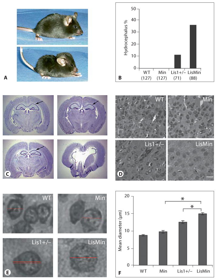
The incidence of hydrocephalus in Lis1+/− mice is increased by a BL/6 genetic background and further by a heterozygous Min mutation. WT = Wild type. A Top panel: a wild-type mouse from a second-generation Lis1+/−/Min backcross. Bottom panel: a compound heterozygote (LisMin) littermate with hydrocephalus. Note the dome-shaped skull and altered posture. B The percentage of offspring with the hydrocephalus phenotype is shown for all offspring obtained during these crosses. The numbers and genotypes are indicated. Parental Black Swiss Lis1+/− mice did not develop hydrocephalus. C Cresyl violet staining of coronal sections of adult brains from littermates (95% BL/6) shows severe disturbances of brain architecture in the LisMin animal (lower right panel). Arrows point out the CA1 region of the hippocampus. D, E Cell bodies in LisMin cortex in these sections appear enlarged. Scale bar = 10 μm. F The somal diameter was determined for 100 cells in digital images (at the same magnification) in the cortex of 3 adjacent brain sections. The diameter was determined by measuring a line through the center of the cells using Axiovision image analysis software (horizontal lines in E). The average somal diameter of neurons in Min mice was not different from that of control mice. Lis1+/− soma were swollen relative to the wild type. This was significantly exacerbated in LisMin brains (* p < 0.00003, Student's t test).