Fig. 5.
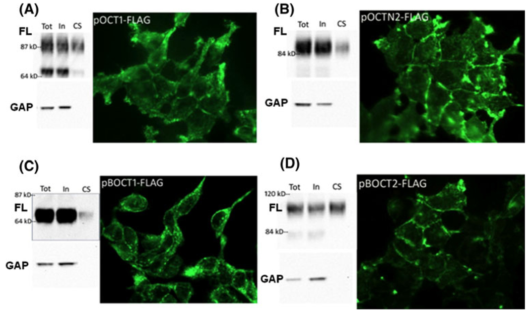
Recombinant BOCT1 and BOCT2 protein is expressed on the cell surface. Stable recombinant HEK cell lines were analyzed for expression of FLAG fusion protein by immunofluorescence and by western blot. a pOCT1-FLAG, b pOCTN2-FLAG, c pBOCT1-FLAG, d pBOCT2-FLAG. Each panel is a composite of the biotinylation experiment (left) and cell staining (right). Left: Cells were incubated with membrane-impermeable sulfobiotin and biotinylated proteins purified on streptavidin beads, followed by western blot analysis using anti-FLAG antibody (upper blots, FL). Each lane contains protein from an equivalent amount of starting material. Tot total protein before purification, In intracellular protein (non-biotinylated), CS cell surface protein (biotinylated). Blots were stripped and incubated with anti-GAPDH to verify that only cell surface protein was purified (lower blots, GAP). Right: Stable transfectants in culture were fixed with paraformaldehyde, permeabilized, and immunostained with anti-FLAG followed by fluorescent secondary antibody

