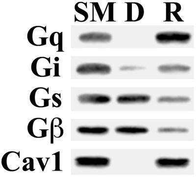
Figure 10.
Dissociation of G proteins from caveolae. Isolated caveolae (V) (20 μg) were diluted in MBS and gently mixed at 4°C for 4 h followed by centrifugation to separate the repelleted caveolar membranes (R) from the soluble phase containing any dissociated proteins (D). An equal volume of starting caveolae (lane SM), R, and D were subjected to Western analysis using antibodies to the indicated proteins.

