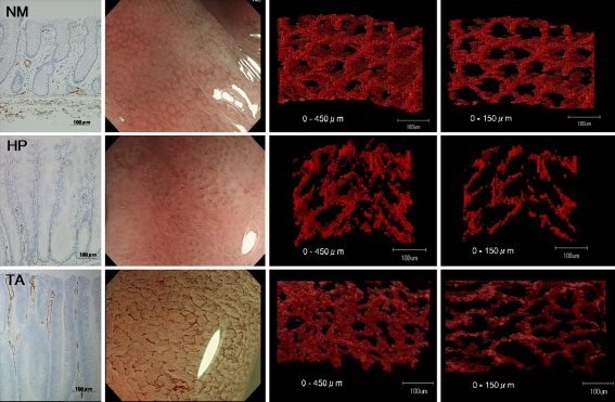
Fig. 1.
CD34-stained sections (first row), images on NBI magnification (second row), and 3-dimensional reconstruction of microvessels (third and fourth rows) in normal mucosa (NM), hyperplasia (HP), and tubular adenoma (TA). Three-dimensional images were reconstructed for microvessels to depths of less than 450 μm (third row) and less than 150 μm (fourth row) from the mucosal surface

