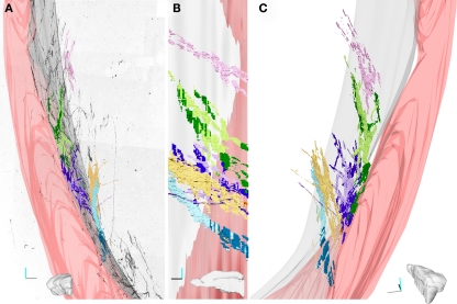
Figure 5.
Three-dimensional reconstruction of four fascicles of DCX+ processes. (A–C). Four fascicles of DCX+ processes were traced in the stack shown in Figure 4. Each fascicles is represented by a different color and its tracts running within the EC are in darker colors. The 3D model was rendered in Blender (www.blender.org). In A the front view of the 3D model is superposed to an inverted image of the MIP of the stack along the z-axis. (B) medial view. (C) Caudal view slightly rotated medially. Only one fascicle (pink) runs exclusively in the striatal parenchyma, while the others run partly inside the EC. The Lilac and green fascicle extend always close to the EC, while the yellow and cyan contact the EC caudally and then turn medially within the striatal parenchyma. The medial surface of the EC is in gray; the surface of a big blood vessel running through the EC is in pink. Orientation Bars: gray line: medial; cyan line: dorsal; dark line: caudal.

