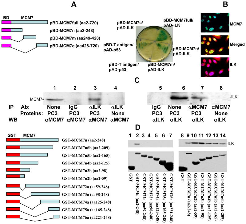
Figure 1. MCM7 binds ILK.
(A) MCM7 binds ILK in Yeast Two Hybrid co-transfection. Left: Schematic diagram of Yeast Two hybrid pGBKT7-MCM7 constructs. Fragments of MCM7 were ligated in frame with the DNA binding (BD) domain of GAL4 protein. Right: Growth and α-galactosidase activity of Yeast AH109 cells co-transfected with the indicated vectors in SD-4 (-leu/-trp/-ade/-his) medium. (B) Co-localization of MCM7 and ILK in PC3 cells. PC3 cells were immunostained with antibodies specific for MCM7 (mouse) and ILK (goat). Immunofluorescence staining was then performed using FITC conjugated antibodies against mouse (MCM7) or Rhodamine-conjugated antibodies against goat (ILK). (C) Co-immunoprecipitation of ILK and MCM7 from PC3 cells. Protein extracts from PC3 cells were immunoprecipitated with the indicated antibodies, electrophoresed in 8% SDS-PAGE and immunoblotted with either anti-MCM7 or ILK antibodies. (D) In vitro binding analysis of GST-MCM7 fusion proteins with ILK. Left: Schematic diagram of GST-MCM7 fusion protein and deletion constructs. Right: Upper panel: Immunoblots of GST-MCM7 fusion proteins pull-down ILK. Lower panel: Coomassie staining of GST-MCM7 fusion proteins.

