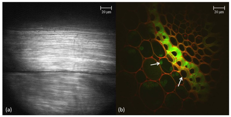
Fig. 6.
(a) An enlarged view of a rat tail tendon optical section taken via SHG with the ultra-slim objective in series with the benchtop system. (b) A false-color image shows Convallaria cells imaged via TPM at two different wavelengths, also with the ultra-slim objective. The left arrow points to a green nucleus and while the right arrow points to a red cell membrane.

