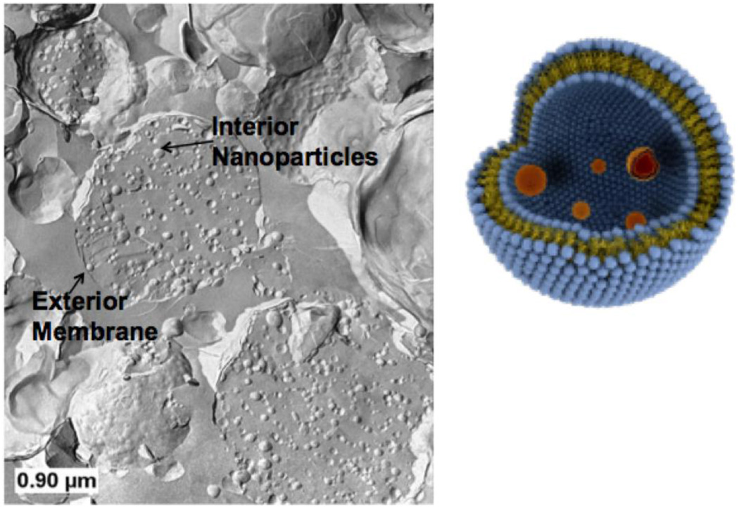
Figure 3.
Schematic (right) and freeze-fracture transmission electron micrograph (left) of vesosome structure. Small, unilamellar vesicles (50 – 100 nm diameter) are encapsulated within 200 – 2000 nm diameter bilayers formed by the transition from the interdigitated LbI phase to the La phase induced by heating the sample above 45° C. Here the interior and exterior compartments are both made of dipalmitoylphosphatidylcholine.

