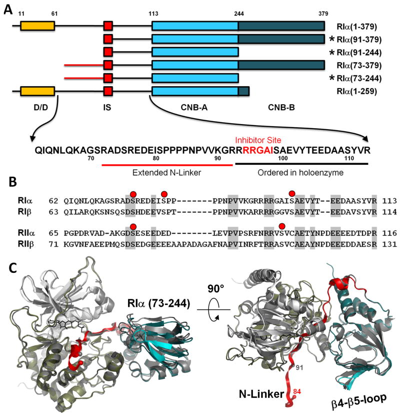
Figure 1. Extended N-Linker in RIα(73-244):C holoenzyme complex.
A. Domain organization of different RIα constructs. Sequence of the linker between Dimerization Domain (D/D) and cAMP binding domain A (CNB-A) is shown. Constructs that were crystallized in holoenzyme complexes are marked with asterisks. B. Sequence alignment of the linkers in different R-subunits. Possible sites of phosphorylation are shown as red dots. C. RIα(73-244):C structure overlaid with the previously solved RIα(91-244):C (dark grey). Catalytic subunit is colored white (N-lobe) and tan (C-lobe). CNB-A is colored teal, the linker is colored red. N-terminal Cα atoms of the RIα constructs are shown as spheres.

