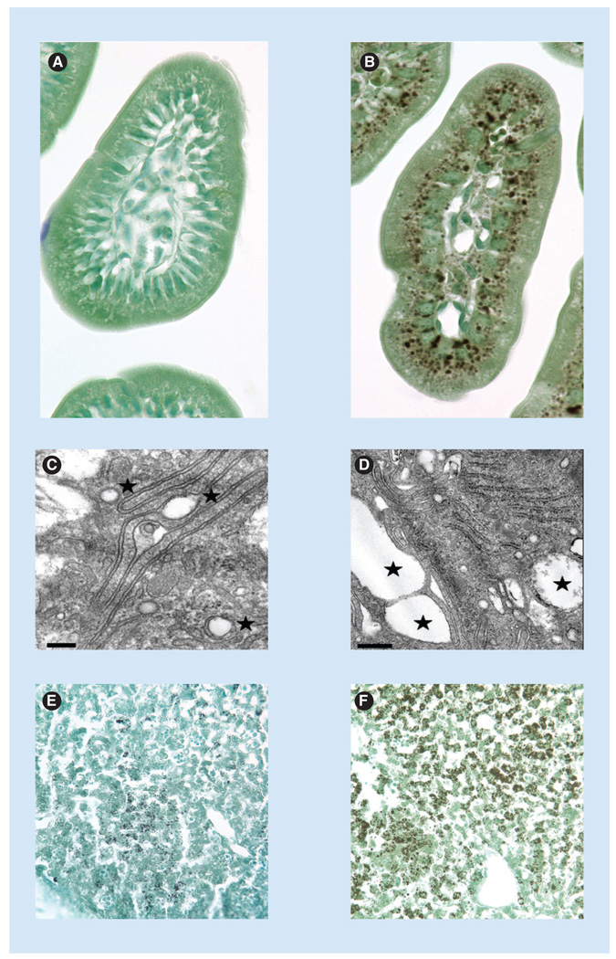
Figure 7. Intestinal and hepatic steatosis in phosphatidylinositol transfer proteins in α-deficient mice.
Intestinal slices stained for neutral lipid content with osmium from (A) pitpα+/+ and (B) pitpα0/0 mice. Note the obvious accumulation of neutral lipid in mutant enterocytes. This accumulation is dependent on nursing and chases only slowly during periods of fast. The phenotype Is also obvious in electron micrographs of the villi of duodenal enterocytes from (C) pitpα+/+ and (D) pitpα0/0 mice (scale bars are 0.2 and 0.5 µM, respectively). Lipid deposits are highlighted by asterisks. Liver slices stained with osmium from (E) pitpα+/+ and (F) pitpα0/0 mice are also shown.

