
Figure 3.
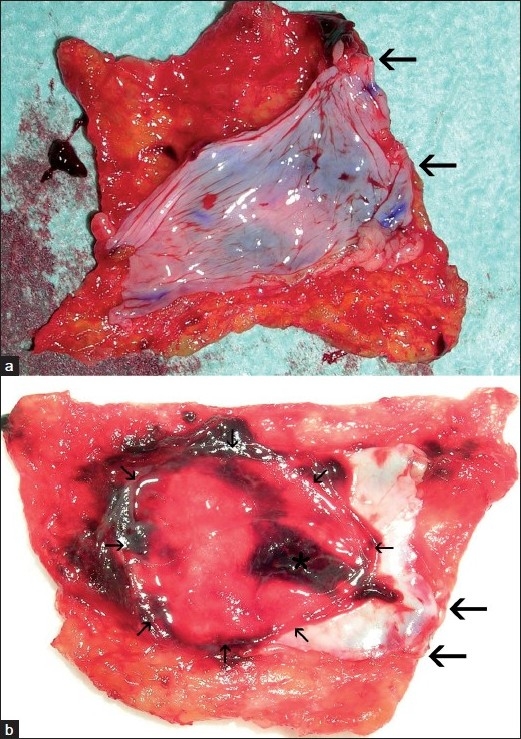
First (panel a) and second (panel b) TachoSil-peritoneal patch harvested from pig number 3 that bled to death. Large arrows demarcate stretches without gluing zones. Small arrows demarcate portion of the peritoneum corresponding to the caval defect. Asterisk marks thrombus